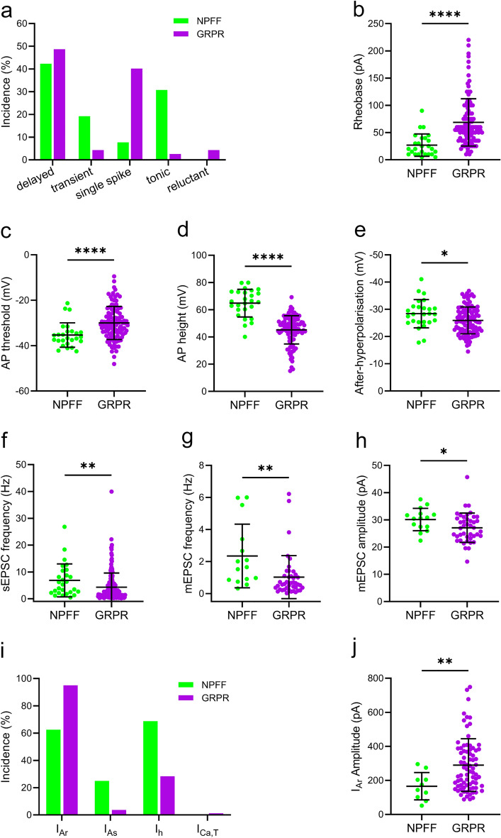
Figure 11.
Comparison of the electrophysiological properties of NPFF cells with those of GRPR cells. Data for the GRPR cells was obtained from Polgár et al18. (a) The incidence of action potential firing patterns differed between NPFF and GRPR cells. NPFF cells exhibited a greater proportion of tonic (30.8 vs. 2.6%) and initial burst firing (19.2 vs. 4.3%) cells and fewer single spike firing cells (7.7 vs. 40.2%). The incidence of delayed firing was similar between both cell types (42.3 vs. 48.7%). NPFF cells were found to have a lower rheobase (26.9 ± 20.5 v.s 68.7 ± 43.4 pA, P < 0.0001, b), action potential voltage threshold (-35.3 ± 5.4 vs. -30.0 ± 7.3 mV, P < 0.0001, c) and a greater action potential height (64.8 ± 10.2 vs. 45.2 ± 10.4 mV, P < 0.0001, d) and after-hyperpolarisation (-28.4 ± 5.2 vs. − 25.9 ± 4.9 mV, P = 0.024, e). In terms of excitatory synaptic input, sEPSC (6.86 ± 6.15 vs. 4.34 ± 5.26 Hz, P = 0.005, f) and mEPSC (2.35 ± 1.99 vs. 1.02 ± 1.34 Hz, P = 0.002, g) frequency in NPFF cells was greater than that in GRPR cells, as was the mEPSC amplitude (30.1 ± 4.1 vs. 27.1 ± 5.4 pA, P = 0.023, h). (i) The incidence of subthreshold voltage-activated currents differed between NPFF and GRPR cells. NPFF cells demonstrated a higher incidence of IAs (25.0 vs. 3.7%) and Ih (68.8 vs. 28.4%), but a lower incidence of IAr (62.5 vs. 95.1%). (j) The peak amplitude of the IAr recorded in NPFF cells was significantly lower than for GRPR cells (165.7 ± 80.3 vs. 289.7 ± 154.7, P = 0.007). All statistical comparisons made using a Mann Whitney test, except for afterhyperpolarisation, which was compared using an unpaired t test.

