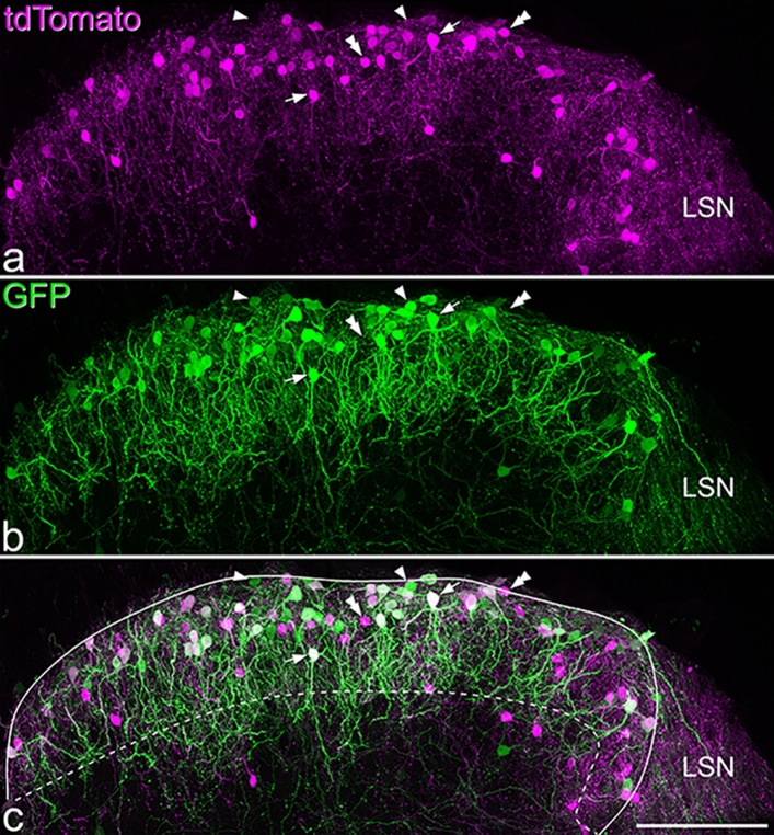
Figure 2.
The distribution of tdTomato- and GFP-labelled cells in the L3 segment of the spinal cord of a NPFFCre;Ai9 mouse that had received intraspinal injections of AAV.flex.GFP. a and b show that both tdTomato- and GFP-positive cells are largely restricted to the superficial dorsal horn. c: the merged image reveals that many cells contain both fluorescent proteins (some indicated with arrows), while some cells are positive for only GFP or tdTomato (some of these are marked with single or double arrowheads, respectively). Dendritic labelling is more prominent in the GFP image, and dendrites of these cells commonly project ventrally into the deeper regions of the dorsal horn. Some labelling with each fluorescent protein is seen in the lateral spinal nucleus (LSN), but labelled cell bodies are seldom seen here. We found that 48% of labelled cells contained both fluorescent proteins, 30% only contained GFP and 22% contained only tdTomato. The solid and dashed lines in c indicate the edge of the grey matter and the approximate border between laminae II and III, respectively. Images are projections of 58 optical sections at 1 μm z-separation. Scale bar = 100 μm.

