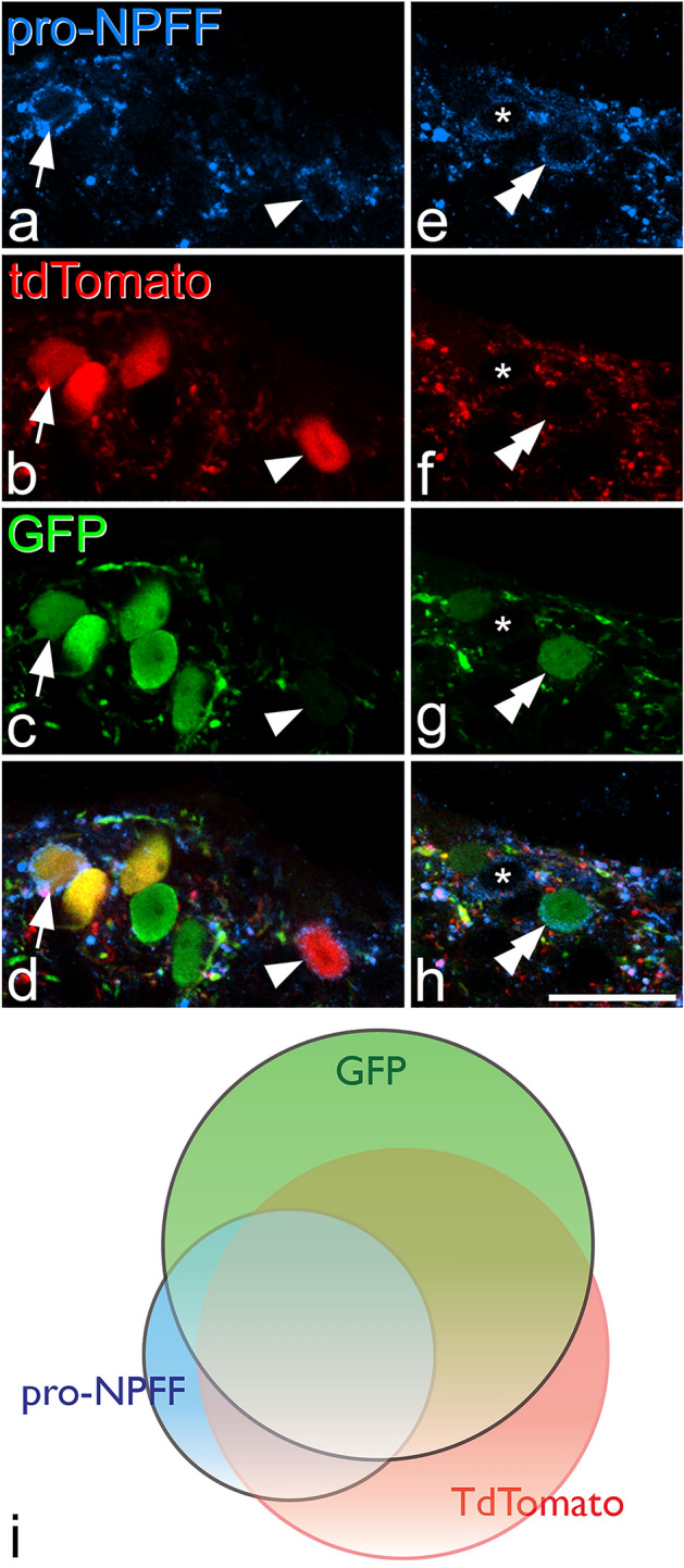
Figure 3.

The relation of pro-NPFF immunoreactivity to expression of tdTomato and GFP in a NPFFCre mouse following intraspinal injection of AAV.flex.GFP. (a)–(d): show part of the section illustrated in Fig. 2 scanned to reveal immunostaining for pro-NPFF (blue) as well as expression of tdTomato (red) and GFP (green). Two pro-NPFF-positive cells can be identified by the presence of immunoreactivity in the perikaryal cytoplasm, which surrounds the unstained nucleus. One of these cells (arrow) contains both fluorescent proteins, while the other (arrowhead) contains tdTomato but lacks GFP. (e)–(h): a nearby field from the same section. Again, this contains two pro-NPFF-positive cells, but in this case one is GFP + /tdTomato-negative (double arrowhead), and the other (asterisk) lacks both fluorescent proteins. Among pro-NPFF-immunoreactive cells, 67% were labelled with both GFP and tdTomato, 7.7% only with GFP, 13.5% only with tdTomato, while 11.9% did not contain either fluorescent protein. All images are projections of 2 optical sections at 1 μm z-separation. (i): Venn diagram showing the extent of overlap of cells labelled with GFP (green), tdTomato (pink) or pro-NPFF-immunoreactivity (blue). Scale bar for (a)–(h) = 10 μm.
