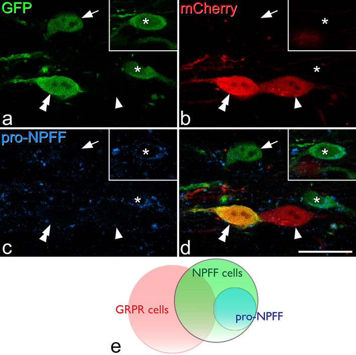
Figure 7.
Immunostaining for pro-NPFF in a section obtained from a NPFFCre;GRPRFlp mouse that had been injected with AAVs coding for Cre-dependent GFP and Flp-dependent mCherry. (a–c) show staining for GFP (green), mCherry (red) and pro-NPFF (blue) in a single confocal optical section, while (d) shows a merged image. The arrow and asterisk mark cells that are positive for GFP and negative for mCherry. The cell indicated with the asterisk contains pro-NPFF, and this is seen more clearly in a different optical section (inset). The cell marked with the arrow lacks pro-NPFF. The cells indicated with single and double arrowheads are both positive for mCherry. One of them (double arrowheads) is also labelled with GFP, while the other (single arrowhead) is not. Both of these cells lack pro-NPFF-immunoreactivity. (e) Venn diagram showing the relationship between cells labelled with each fluorescent protein and pro-NPFF immunoreactivity. pro-NPFF was present in 0.4% of GRPR cells (labelled with mCherry), 25% of NPFF cells (labelled with GFP) and 42% of the cells that were GFP-positive/mCherry-negative. Scale bar = 20 μm.

