Figure 9.
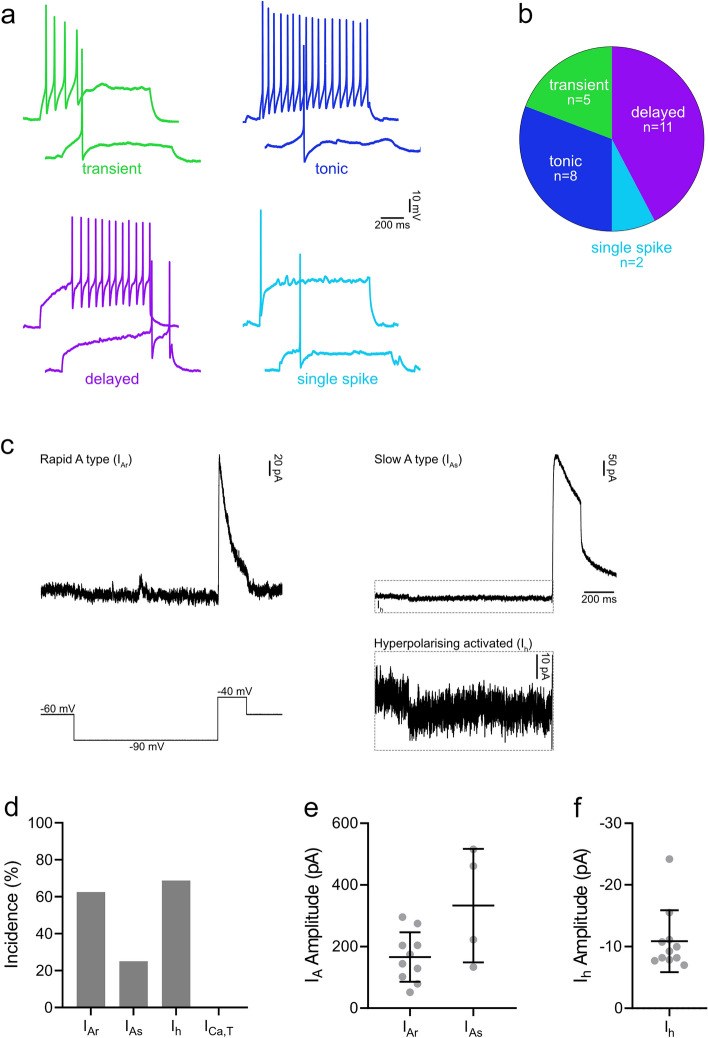
Action potential firing patterns and subthreshold voltage-activated currents in NPFF cells. (a) Examples of action potential firing patterns observed in NPFF cells in response to 1 s suprathreshold current injections. (b) The most prominent type of firing pattern seen in NPFF cells was delayed firing (11/26; 42.3%), with smaller proportions exhibiting tonic (8/26; 30.8%), transient (5/26: 19.2%) or single spike firing (2/26: 7.7%). (c) Representative traces demonstrating the subthreshold voltage-activated currents in NPFF cells that were revealed using a voltage step protocol that hyperpolarised cells from -60 to -90 mV for 1 s and then depolarised cells to -40 mV for 200 ms (bottom left trace). The currents that are revealed by this protocol were classified as rapid (IAr) or slow (IAs) A-type potassium currents, or hyperpolarising-activated currents (Ih). The example traces of IAr (top left) and IAs with Ih (top right) show an average of 5 traces. The example of Ih (dashed outline) is shown at a different y-axis scale (bottom right). (d) Almost all NPFF cells displayed IA, which was mostly classified as IAr (10/16; 62.5%), but with some exhibiting IAs (4/16; 25.0%). Many cells exhibited Ih (11/16; 68.8%), which was typically seen in addition to IAr (5/16; 31.3%) or IAs (4/16; 25.0%). The peak amplitude of IA was 165.7 ± 80.3 and 333.0 ± 184.1 pA for IAr and IAs, respectively (e) and the amplitude of Ih, measured during the final 200 ms of the hyperpolarising step, was -10.9 ± 5.0 pA (f).

