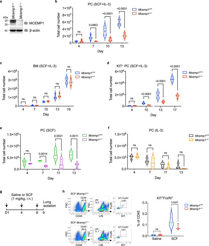
Fig. 2. MCEMP1 deficiency impairs SCF-induced mast cell proliferation.
a Loss of MCEMP1 protein expression in Mcemp1–/– mice. Lungs were immunoblotted with MCEMP1 antibody. Blots are representative of two independent experiments. b, c Cell growth kinetics of Mcemp1+/+ and Mcemp1–/– peritoneal cell (PC, n = 5 per group) or bone marrow cells (BM, n = 4 per group) cultured with SCF and IL-3 for the indicated days. d Cell growth kinetics of Mcemp1+/+ and Mcemp1–/– KIT-positive PC isolated by magnetic beads and cultured with SCF and IL-3 for the indicated days. e, f Absolute counts of Mcemp1+/+ and Mcemp1–/– PC cultured with either SCF or IL-3 for the indicated days (n = 3 per group). g Schematics of SCF intranasal (i.n.) challenge of Mcemp1+/+ and Mcemp1–/– mice. h Representative flow cytometry plots illustrating the gating strategy to identify KIT/FcεRI double-positive mast cells; CD45+LIN–KIT+FcεRI+. The percentages of KIT+FcεRI+ mast cells in the lungs of Mcemp1+/+ or Mcemp1–/– mice challenged with saline or SCF (n = 6-7 mice per group). Data are presented as violin plot with lines at median and quartiles and p-values were determined by two-way ANOVA with Sidak’s multiple comparison in b, c, d, e, f, h. ns, not significant.

