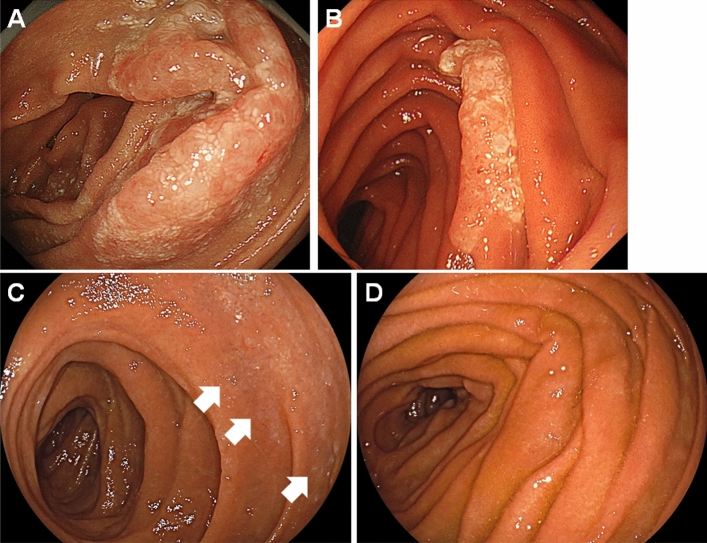
Figure 2.
Representative endoscopic images of a case showing spontaneous regression of duodenal follicular lymphoma. A duodenal lesion showing whitish villi was detected in a 63-year-old woman (A). The area of the whitish lesion reduced 4 years after the diagnosis (B). The whitish lesion became faint 6 years after the diagnosis (C). The duodenal lesion disappeared and no lymphoma cells were detected on biopsied specimen 7 years after the diagnosis (D).

