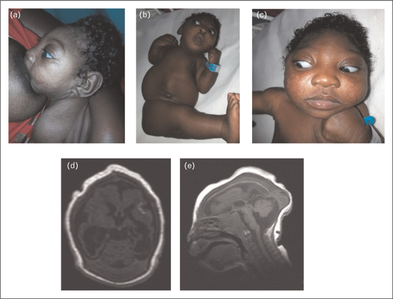
FIGURE 2.
Panels (a, b, c above) show female infant at age 6 weeks (sucking at mother's breast), showing severe microcephaly, sloping fore-head, facial disproportion with ‘over-sized’ facial features, appearance of proptosis, horizontal nystagmus with bilateral optic atrophy. Infant also displays clenched upper limbs with cortical fisting, diastasis of ‘recti abdomini’ muscles, severe arthrogryposis and ‘rocker bottom’ feet. Panels (d and e, above) reveal infant's MRI of the skull and brain displaying marked microcephaly, collapsed skull bones with extensive scalp folding. There is decreased hemispheric parenchymal volume loss with decreased salvation and evidence of calcification (d). There are septations in the occipital horns of the lateral ventricles as well as evidence of a vermian hypoplasia, in keeping with a Dandy Walker variant. Mother gave signed, written, informed consent with her permission for these photographs to be used for the purposes of medical education, publication and research.

