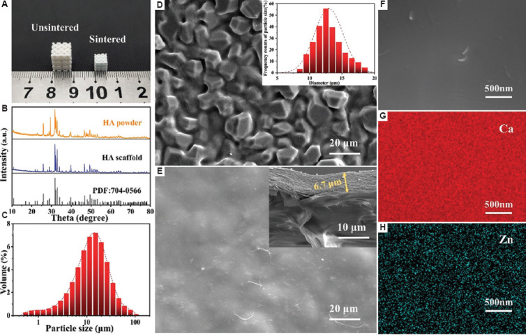
Figure 3.

(A) Images of unsintered and sintered hydroxyapatite (HA) scaffolds. (B) X-ray diffraction spectra of HA powder and scaffold. (C) Particle size distribution of ceramic powders. (D) Scanning electron microscope (SEM) image of HA scaffold; the inset image shows the size distribution of ceramic particle on the scaffold surface. (E) SEM image of HA scaffold with coatings; the inset image shows the cross section of coatings. (F) High-magnification SEM image of coatings. (G and H) EDS mapping of Ca and Zn on CHA-H sample.
