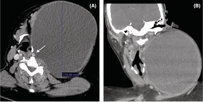
FIGURE 1.

On axial view, the preoperative computed tomography (CT) imaging of the branchial cleft cyst shows a cystic mass measuring greater than 17 centimeters in longest dimension with associated left common carotid artery compression (white arrow) without stenosis relative to contralateral carotid artery (A). Coronal CT view shows rightward tracheal and parapharyngeal deviation secondary to mass effect (B).
