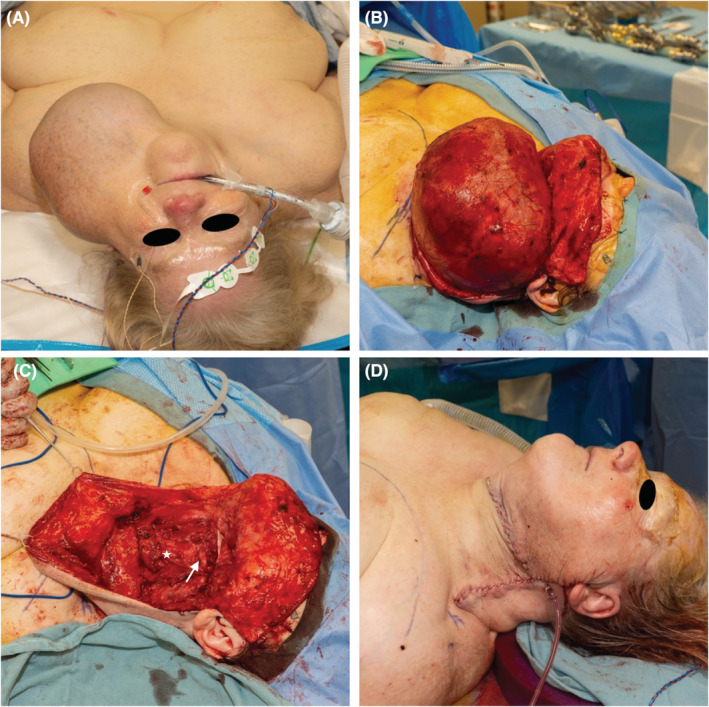
FIGURE 2.

The preoperative exam reveals findings of a massive left neck cyst (A). The in situ preserved cyst wall are noted after raising subplatysmal flaps (B). After cyst excision, the common carotid artery (white star) and hypoglossal nerve (white arrow) are visualized (C). Excess skin and subcutaneous fat is resected after excision of the cyst (D).
