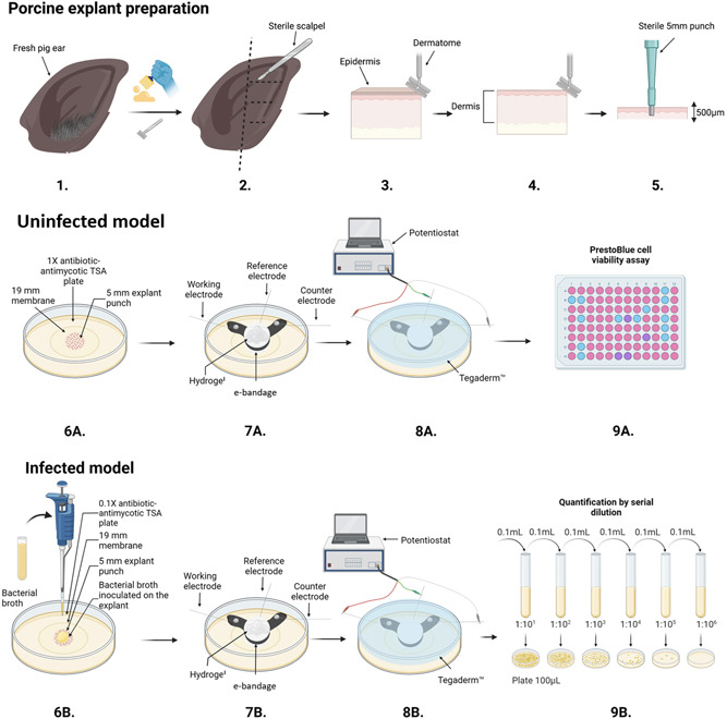
Figure 1.

Schematic of porcine explant preparation (1–5), uninfected model (6A–9A), infected model experiments (6B–9B), e‐bandage placement and connection to potentiostat (7–8), PrestoBlue cell viability assay (9A), serial dilution and CFU quantification (9B). The figure was prepared using BioRender©, and modified from (Tibbits et al., 2022).
