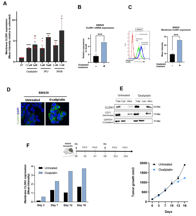
Fig. 2.
CLDN1 is overexpressed at the membrane of colorectal cancer cells after exposure to chemotherapy drugs. (A) Flow cytometry analysis of CLDN1 expression at the surface of SW620 cells incubated with conventional chemotherapy agents (5-FU, SN38, oxaliplatin) at two concentrations. * p ≤ 0.05, ** p ≤ 0.01, *** p ≤ 0.001, **** p ≤ 0.0001 (Student’s t-test). (B) Relative CLDN1 gene expression in SW620 cells before (-) and after (+) incubation with oxaliplatin (1.2 µM for 72 h); *** p ≤ 0.001 (Student’s t-test). (C) Membrane CLDN1 expression analyzed by flow cytometry in SW620 cells before (-) and after (+) incubation with oxaliplatin (1.2 µM for 72 h); *** p ≤ 0.001 (Student’s t-test). (D) Immunofluorescence analysis of CLDN1 membrane expression in SW620 cells before (-) and after (+) incubation with oxaliplatin (1.2 µM for 72 h) (E) Subcellular localization of CLDN1 by western blotting in SW620 cells before (-) and after (+) incubation with oxaliplatin (1.2 µM for 72 h). CD71 and β-actin were used as markers for the membrane and cytoplasm fractions, respectively. Cyto, cytoplasm; Mbm, membrane. (F) In vivo kinetics of CLDN1 expression at the tumor cell membrane. Top: experimental setup: mice xenografted with SW620 cells were treated at day 0 and day 9 or not with oxaliplatin (Ox; 2 mg/kg). At the indicated time points, two tumors per condition were removed, dissociated and analyzed by flow cytometry. Left: Histograms showing CLDN1 membrane expression (g-mean) in untreated (black) and oxaliplatin-treated (blue) tumors. Right: tumor growth curves in untreated and oxaliplatin-treated mice xenografted with SW620 cells

