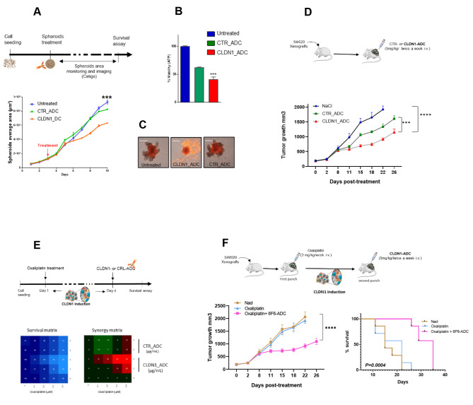
Fig. 5.
Therapeutic effect of an anti-CLDN1 ADC in colorectal cancer cells and “one-two punch” therapeutic approach. (A) Growth curve of SW620 cell spheroids incubated with 10 µg/mL of 6F6-ADC, Control-ADC (anti-CD20 mAb), or not for 7 days monitored with the Celigo™ imaging cytometer. (B) Cell survival in spheroids at the experiment end was determined with a cytotoxicity assay. The luminescence intensity (i.e. viable cells) was measured and compared in the three conditions described in (A). (C) Spheroids were incubated with 1 µg/m of propidium iodide (PI) that emits a red fluorescence red when incorporated in dead cells. Images were acquired using the Celigo™ imaging cytometer. (D) Effect of 6F6–ADC and control-ADC (Human IgG1, kappa Isotype Control) on the growth of SW620 cell xenografts in athymic nude mice. Mice were treated or not (blue) with 5 mg/kg of Control-ADC (green) or 6F6-ADC (red) per week starting when tumors reached 100 mm3 (n = 7 mice per group). (E) In vitro analysis of the effects of the oxaliplatin + 6F6-ADC combination in SW620 cells. At 24 h post-seeding cells were incubated with increasing doses of oxaliplatin and 72 h later with increasing doses of 6F6-ADC or control-ADC. One week later, the cell viability assay was performed. The blue matrix represents cell viability. In the synergy matrix, red, black, and green represent synergy, additivity, and antagonism, respectively. (F) Tumor growth curves in mice xenografted with SW620 cells untreated, treated with oxaliplatin alone or with the sequential combination of oxaliplatin and 6F6-ADC (7 mice per group). Adapted Kaplan-Meier curves using the time taken to reach a tumor volume of 1500 mm3 in untreated, oxaliplatin-treated and sequential combination-treated mice (log-rank test)

