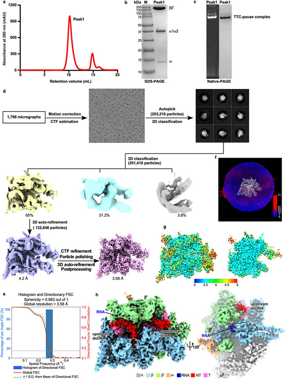
Extended Data Fig. 1. The processing pipeline for single-particle reconstitution of E. coli TTC-pause.
a. Elution peaks of TTC-pause (peak1) from a size-exclusion column. b. The SDS-PAGE and c. the native-PAGE of peak 1. The gel was first stained with SYBR Gold for nucleic acids and then with Coomassie Brilliant Blue for proteins. The experiment has been repeated three times with similar results. d. The flowchart of data processing of TTC-pause. e. The 3D FSC plot. The dotted line represents 0.143 cutoff of the global FSC curve. f. The angular distribution of single-particle projections by number of particles of each projection. g. The cryo-EM map of TTC-pause colored by local resolution. h. The cryo-EM map of TTC-pause in front and side views.

