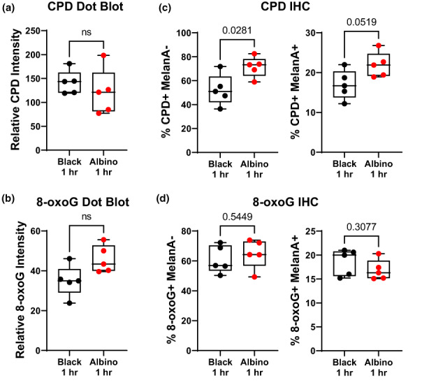
FIGURE 4.

Cell‐intrinsic melanin does not enhance initial UV damage in melanocytes. (a, b) Box and whisker plots of CPD (“a”) and 8‐oxoG (“b”) staining intensity in dot blots of DNA isolated from the dorsal skin of black and albino TpN mice 1 h after UV irradiation. (c, d) Quantification of immunofluorescent co‐staining of CPD (“c”) or 8‐oxoG (“d”) and MelanA (melanocyte maker) in formalin‐fixed skin sections from black and albino TpN mice 1 h after UV irradiation. (a–d) Dots represent biological replicates. Boxes span the 25th–75th percentile with whiskers from the minimum to maximum value. Statistical significance was determined using Welch's t‐tests.
