Abstract
Introduction:
Bilateral neck of femur stress fractures is rare clinical presentation in elderly population. Diagnosis of such fractures when presented can be difficult with inconclusive radiographs and hence their early diagnosis by high index of suspicion and their management can avoid further complications in this age group. In this case series, we report three elderly patients with different predisposing factors for their fractures and treatment options chosen with detailed discussion about the management.
Case Report:
These case series of three elderly patients with bilateral neck of femur fracture were associated with different predisposing factors. Grave’s disease or primary thyrotoxicosis, steroid induced osteoporosis, and renal osteodystrophy were the identified risk factors in these patients. Biochemical evaluation for osteoporosis in these patients revealed significant derangement in levels of vitamin D, Alkaline phosphatase, and serum calcium. One of these patients was operated with hemiarthroplasty and osteosynthesis with percutaneous screw fixation on the other side. Management of osteoporosis, dietary modifications, and lifestyle changes in these patients also had significant impact on their prognosis.
Conclusion:
Stress fractures occurring in elderly individuals with simultaneous bilateral presentation are rare and can be prevented if taken care on their risk factors. As radiographs remain inconclusive few times in these kinds of fracture, a high degree of suspicion is to be kept in mind. With advanced diagnostic tools and surgeries, they carry good prognosis if timely intervention is provided.
Keywords: Pathological, neck of femur fracture, bilateral, geriatric
Learning Point of the Article:
Bilateral stress neck of femur fractures in in elderly carries good prognosis if timely intervention is provided.
Introduction
Stress fracture is produced on a bone that has not been adapted to the prolonged and repetitive muscular activity. Stress fractures may be presented as fatigue fractures occurring due to the abnormal stress to a bone with normal characteristics and on the other hand or as insufficiency fractures occurring due to normal stress on a bone with decreased mineral or elastic resistance [1]. The pathophysiologic outcome is the combination of an imbalance between bone remodeling capacity and repetitive stress. Endocrine, metabolic, and pharmacologic causes have been found.
Distal shaft, proximal shaft, and femoral neck are the three femoral regions which are more susceptible to stress fracture [2, 3, 4]. Stress fractures occurring at the femoral neck represent 5% of all stress fractures in the general population, but in elderly patients, it is 16% [5, 6]. There are various risk factors involved in causing stress fracture out of them Senile and post-menopausal osteoporosis contributed the most [7]. Unilateral stress fracture of neck femur in elderly population is common presentation, but bilateral fracture either simultaneous or sequential is rare and needs high index of suspicion and proper evaluation for their diagnosis and causes [8].
Here, we are reporting a series of three cases of stress fracture of bilateral neck of femur in elderly population with different causes, along with a detailed literature review.
Case Report
Case 1
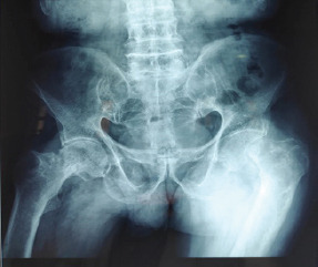
A 70-year-old male with Grave’s disease presented with severe pain in both hips region and difficulty while walking since last 1 month without any associated night pain, trauma and constitutional sign and symptoms. The patient was investigated and evaluated in form of in form of deficient levels of serum vitamin D (23 ng/ml), elevated levels of free thyroxin hormones with low levels of TSH (<0.01 microIU/ml), with normal levels of serum calcium, phosphate, and ALP with negative inflammatory markers. Plane radiographs were suggestive of stress fracture of bilateral neck of femur in Fig. 1. On confirmation of Grave’s disease with anti-TPO antibody testing, the patient was started on Tablet Carbimazole 10 mg and Tablet Propranolol 10 mg daily and the patient refused for proposed surgery.
Figure 1.

Bilateral simultaneous fracture neck of femur of Case 1.
Case 2
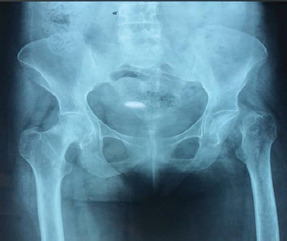
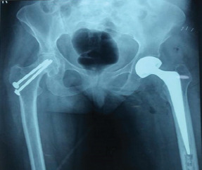
A 50-year-old female with rheumatoid arthritis taking steroids for long time presented with pain in the left hip and limping for 6 months and also developed pain in opposite hip for 1 month. Radiographs showed undisplaced fracture of the right neck femur and displaced and resorbed fracture neck on the left side, as shown in Fig. 2. On investigating, her serum levels of calcium and phosphate were normal and vitamin D levels were low (16 ng/ml) and quantitative rheumatoid factor (305 IU/mL) and anti-CCP antibody levels (698 IU/mL) were too high with elevated levels of erythrocyte sedimentation rate (ESR) and CRP levels. Following pre-anesthetic fitness, hemiarthroplasty of the left hip and in situ fixation with two partially threaded cancellous screws was done, as shown in Fig. 3.
Figure 2.

Undisplaced fracture of the right side neck of femur and displaced and resorption of fracture of neck of femur left side in case 2.
Figure 3.

Post-operative radiograph showing in situ cancellous screw fixation for the right side and hemiarthroplasty for the left hip in case 2.
Case 3
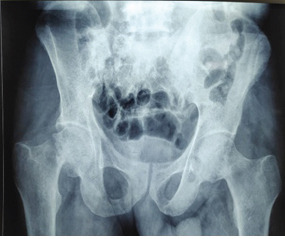
A 80-year-old male presented with inability to bear weight and pain in bilateral hip since 2 weeks with no prior history of trauma. On evaluation, the patient was a known case of diabetes mellitus and hypertension with chronic kidney disease with deficient serum levels of vitamin D (6 ng/ml), calcium (6.3 mg/dl), and elevated phosphate (6.2 mg/dl) and elevated serum creatinine level (3.2 mg/dl). Radiographs revealed simultaneous bilateral neck of femur fracture, as shown in Fig. 4. Eventually, the patient developed urosepsis with multiple organ dysfunction syndrome (MODS) and expired.
Figure 4.

Radiograph showing bilateral stress fracture of femur neck in Case 3 with chronic renal failure.
Discussion
Stress fractures may develop in up to 5% of runners and military trainers [9]. Of those patients who develop stress fractures, about 3–8% of the fractures are in the femoral neck being subjected to excess force while walking. In elderly patients, 16% of stress fractures occur in femoral neck [10, 11]. As most of neck stress fracture reported in the literature occur in young adults, especially military trainers and athletes, incidence of neck stress fracture in elderly people is not well known.
Just like any other preventable disease, stress fracture can also be prevented with knowledge on their risk factors. Non-modifiable risk factors play a significant role such as white race, female sex, menopause, high bone turnover, low bone density, and inadequate muscle function [12]. Modifiable risk factors include overtraining, vitamin D insufficiency, energetic nutrition deficit, nicotine and alcohol abuse, steroid use, low adult weight, anorexia, or bisphosphonate therapy [13, 14, 15]. Varies cofactor coexistence make it difficult for isolation of an etiological factor [16]. Inadequate diet and nutrition also contribute in development of stress fractures in third world countries.
In our case reports, the cause for stress fracture was found to be Graves disease in case 1, though uncommon in this age patient, which had less prominent classic symptoms and signs of hyperthyroidism, diagnosis was made due to high index of suspicion. There are fewer literature of Grave’s disease presenting with bone demineralization and pathological fracture. We believe that our case 2, with rheumatoid arthritis and associated deformities, had osteoporotic bone due to chronic steroid intake and in case 3, it was renal osteodystrophy secondary to chronic renal failure which made the bones osteoporotic and susceptible to simultaneous bilateral neck of femur stress fracture. The biochemical evaluation reports in these patients have been detailed in Table 1.
Table 1.
Detailed investigation of the patients
Fullerton and Snowdy [12] classification of femur neck stress fracture comprising three categories as first category, tension-side of the femoral neck fractures, where fracture is placed in the superior part of the femoral neck while in second category compression-side stress fractured, where the fracture line is seen in the inferior aspect of femoral neck and more common than the previous category. In the third category, fracture is already displaced in the initial X-ray, irrespective whether the fracture started initially in the tension or compression side.
AS per experience, we recommend to evaluate these patients in form of blood investigations such as PTH and serum Vitamin D levels to rule out insufficiency fractures. While suspecting malignancy ESR, liver functions, and renal functions plat a key role malignancy and serum protein electrophoresis is mandatory in multiple myeloma suspicion. Radiological investigation such as complete skeletal survey plays a key role in diagnosing occult stress fracture. More comprehensive investigation like a PET scan help to look for possible sites of primary malignancy or other sites of involvement. In case, when X-ray findings are inconclusive MRI play a major role mostly in non-ambulatory patients, although costly but delay in diagnosis of fractures of weight-bearing can be avoided which can lead to immobility and its complications. The definitive treatment of the stress fracture can be decided by the presence of fracture displacement or avascular necrosis. In such cases, surgery with hip arthroplasty can be indicated. When an early diagnosis is made, and the fracture is undisplaced, osteosynthesis may be considered either with partially threaded cannulated screws or cephalomedullary nailing and if fractures are incomplete and the patient is unfit for surgery conservative trial can be advised. Moreover, the same protocol being followed in our case report has given a good outcome and patient satisfaction.
Conclusion
Bilateral femoral neck stress fracture in geriatric population is uncommon. In elderly patients, it is usually associated with multiple factors such as vitamin D deficiency, osteoporosis, metabolic bone disease, renal abnormalities, steroid use, and malignancy. Hence, complete evaluation of each and every case is required. In the above-mentioned series, we had presented three cases of bilateral fracture neck of femur associated with steroid use in rheumatoid arthritis in first case, due to Grave’s disease in second and renal failure secondary to diabetes mellitus in third case. Management of these patient requires a multidisciplinary approach, it depends on the type of fracture and general condition of patient. Educating the geriatric population with lifestyle changes such as avoidance of trauma and other stressful activities, dietary modifications to include Vitamin D rich diet and diet fortified with calcium, definitely large number of stress fractures, and their untoward consequences in this age group can be avoided.
Clinical Message.
Stress fractures occurring in elderly individuals with simultaneous bilateral presentation are rare and can be prevented if taken care on their risk factors. Complete radiological and biochemical evaluation to be done in all such patients. Diagnosis of these fractures needs high index of suspicion as radiographs remain inconclusive few times. With advanced diagnostic tools and surgeries, they carry good prognosis if timely intervention is provided.
Biography


Footnotes
Conflict of Interest: Nil
Source of Support: Nil
Consent: The authors confirm that informed consent was obtained from the patient for publication of this case report
References
- 1.Egol KA, Koval KJ, Kummer F, Frankel VH. Stress fractures of the femoral neck. Clin Orthop Relat Res. 1998;348:72–8. [PubMed] [Google Scholar]
- 2.Niva MH, Kiuru MJ, Haataja R, Pihlajamaki HK. Fatigue injuries of the femur. J Bone Joint Surg Br. 2005;87:1385–90. doi: 10.1302/0301-620X.87B10.16666. [DOI] [PubMed] [Google Scholar]
- 3.Korpelainen R, Orava S, Karpakka J, Siira P, Hulkko A. Risk factors for recurrent stress fractures in athletes. Am J Sports Med. 2001;29:304–10. doi: 10.1177/03635465010290030901. [DOI] [PubMed] [Google Scholar]
- 4.Fredericson M, Jennings F, Beaulieu C, Matheson GO. Stress fractures in athletes. Top Magn Reson Imaging. 2006;17:309–25. doi: 10.1097/RMR.0b013e3180421c8c. [DOI] [PubMed] [Google Scholar]
- 5.Pentecost RL, Murray RA, Brindley HH. Fatigue, insufficiency, and pathologic fractures. JAMA. 1964;187:1001–4. doi: 10.1001/jama.1964.03060260029006. [DOI] [PubMed] [Google Scholar]
- 6.Markey KL. Stress fractures. Clin Sports Med. 1987;6:405–25. [PubMed] [Google Scholar]
- 7.Gedmintas L, Solomon DH, Kim SC. Bisphosphonates and risk of subtrochanteric, femoral shaft, and atypical femur fracture:A systematic review and meta-analysis. J Bone Miner Res. 2013;28:1729–37. doi: 10.1002/jbmr.1893. [DOI] [PMC free article] [PubMed] [Google Scholar]
- 8.Clohisy JC, Carlisle JC, Sierra RJ, Beaulé PE, Kim YJ, Trousdale RT, et al. A systematic approach to the plain radiographic evaluation of the young adult hip. J Bone Joint Surg Am. 2008;90:47–66. doi: 10.2106/JBJS.H.00756. [DOI] [PMC free article] [PubMed] [Google Scholar]
- 9.Amstrong DW, Rue JP, Wilckens JH, Frsassica FJ. Stress fracture injury in young military men and women. Bone. 2004;35:803–16. doi: 10.1016/j.bone.2004.05.014. [DOI] [PubMed] [Google Scholar]
- 10.Pihlajamäki HK, Ruohola JP, Weckström M, Kiuru MJ, Visuri TI. Long-term outcome of undisplaced fatigue fractures of the femoral neck in young male adults. J Bone Joint Surg Br. 2006;88:1574–9. doi: 10.1302/0301-620X.88B12.17996. [DOI] [PubMed] [Google Scholar]
- 11.Carpintero P, Berral FJ, Baena P, Garcia-Frasquet A, Lancho JL. Delayed diagnosis of fatigue fractures in the elderly. Am J Sports Med. 1997;25:659–62. doi: 10.1177/036354659702500512. [DOI] [PubMed] [Google Scholar]
- 12.Fullerton LR, Jr, Snowdy HA. Femoral neck stress fractures. Am J Sports Med. 1988;16:365–77. doi: 10.1177/036354658801600411. [DOI] [PubMed] [Google Scholar]
- 13.Breer S, Krause M, Marshall RP, Oheim R, Amling M, Barvencik F. Stress fractures in elderly patients. Int Orthop. 2012;36:2581–7. doi: 10.1007/s00264-012-1708-1. [DOI] [PMC free article] [PubMed] [Google Scholar]
- 14.Koh JS, Goh SK, Png MA, Kwek EB, Howe TS. Femoral cortical stress lesions in long-term bisphosphonate therapy:A herald of impending fracture? J Orthop Trauma. 2010;24:75–81. doi: 10.1097/BOT.0b013e3181b6499b. [DOI] [PubMed] [Google Scholar]
- 15.Isaacs JD, Shidiak L, Harris IA, Szomor ZL. Femoral insufficiency fractures associated with prolonged bisphosphonate therapy. Clin Orthop Relat Res. 2010;468:3384–92. doi: 10.1007/s11999-010-1535-x. [DOI] [PMC free article] [PubMed] [Google Scholar]
- 16.Chihaoui MB, Elleuch M, Sahli H, Cheour I, Romdhane RH, Sellami S. Stress fracture:Epidemiology, physiopathology and risk factors. Tunis Med. 2008;86:1031–5. [PubMed] [Google Scholar]


