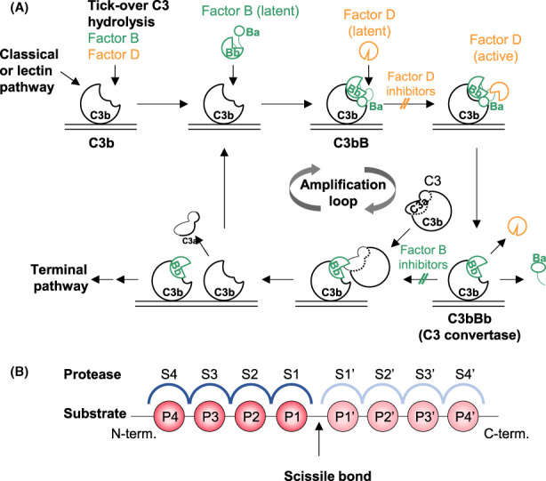
FIGURE 1.

Protease function in the alternative pathway. (A) Proteolytic activation of the alternative complement pathway. For detailed description see text. (B) Schematic representation of the active site of proteases, indicating individual binding pockets of the protease (S, blue) and corresponding substrate amino acids (P, red), N‐terminally (non‐prime, dark colors) or C‐terminally (prime, light colors) of the scissile bond
