
FIGURE 1.
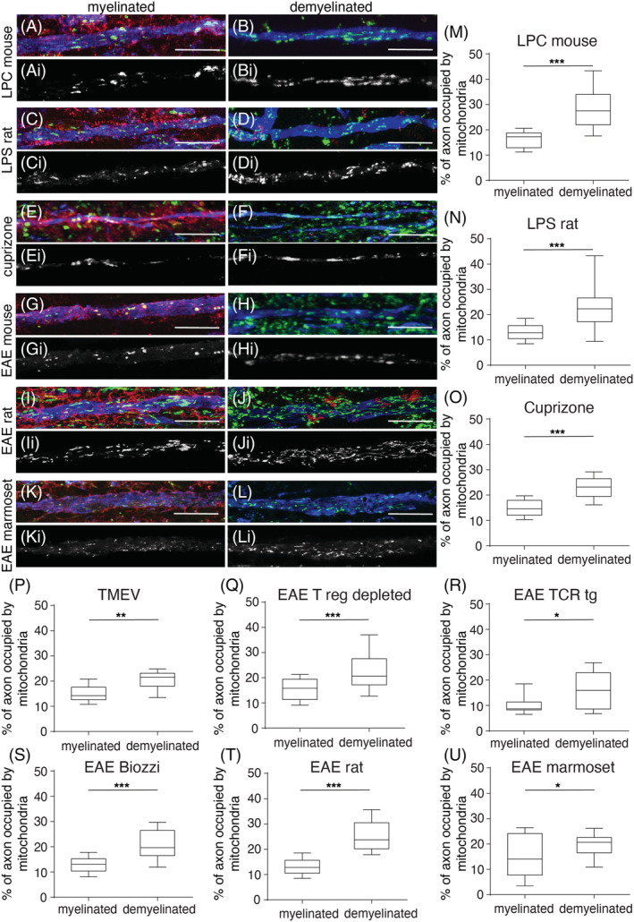
Axonal mitochondrial content consistently increases upon experimental demyelination. Compared with myelinated axons from controls, in triple labelled immunofluorescent confocal images (column to the left with MBP in red, neurofilament‐H in blue and porin in green), mitochondria are more prevalent in demyelinated axons (column to the right) from all the models. The greyscale images (Ai‐li) show porin‐positive elements within axons from the corresponding triple labelled colour images (A‐L). The quantitation of axonal mitochondrial content shows a significant increase in the lysolecithin‐induced focal lesions (LPC, A‐B and M), lipopolysaccharide‐induced focal lesions (LPS, C‐D and N), cuprizone model (E‐F and O), Theiler's murine encephalomyelitis virus (TMEV) model (P) as well as experimental autoimmune encephalitis (EAE, G‐L and Q‐U) in mice (C57BL6, SJL/J and Biozzi ABH), rat (dark agouti) and marmoset species (the area of porin‐positive elements as a percentage of axon area); 20 axons per region were randomly selected from each animal for quantitation. The box plots indicate the median, inter‐quartile range (25%–75%) and 90% confidence interval. *p < 0.05, **p < 0.01 and ***p < 0.01. The scale bar indicates 10 μm.