
Figure 3.
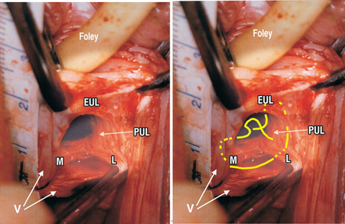
Live anatomy—surgical binding of loose pubourethral ligaments (PUL). Left: Original live anatomical dissection 6 of PUL (left incision) during a two incision IVS operation. 2 The tape measure overlies the urethra. The left paraurethral sulcus has been incised along its length and opened out laterally with forceps. External urethral ligament (EUL) is the EUL which sits in front of the pubic symphysis (PS). The PUL, originates behind PS from its lower posterior part. Coming down from PS, PUL splits into two parts, medial (M) to insert into the side of the midurethra and L (lateral). “L” attaches laterally to pubococcygeus muscle (not seen) then comes down to attach to the vagina (V). Right: No.2 or preferably, No.5 polyester sutures bind both branches of PUL to fascias attached to pubic bone, urethra, vagina and pubococcygeus muscle (PCM), essentially as performed in the original operation. 1