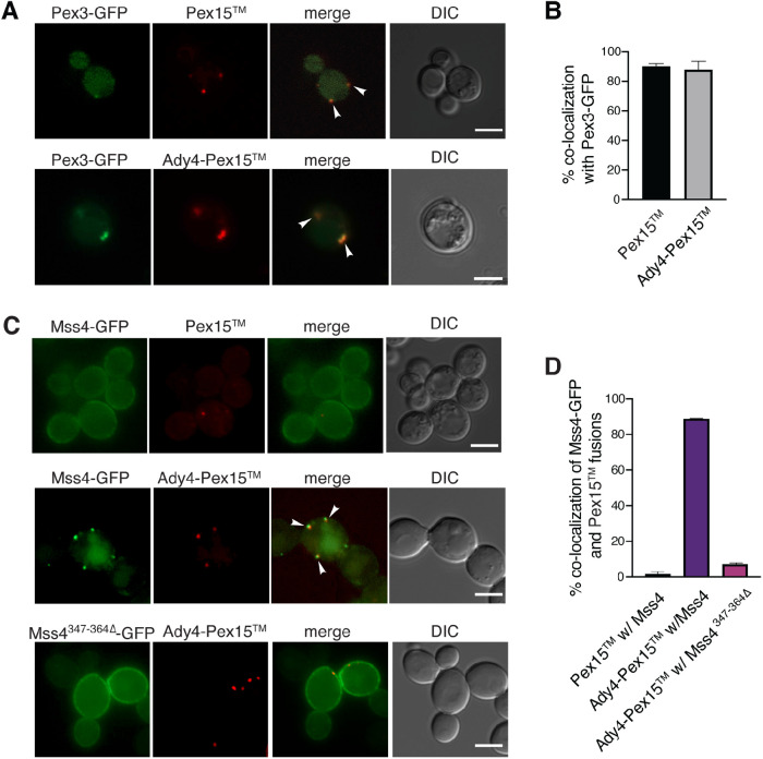
FIGURE 4:
Ady4 can target Mss4 to an ectopic location in vegetative WT cells. (A) A WT diploid (AN120) was transformed with plasmids expressing PEX3-eGFP (pRS424-PTEF1Pex3-eGFP) and PEX15 (pRS316-PTEF1-mOrangePex15) or ADY4- PEX15 (pRS426-PTEF1Ady4-mOrange-Pex15) and localization of the fusion proteins was examined by fluorescence microcopy. Arrowheads indicate colocalization of Pex3 and the Pex15 fusions. Scale bars = 5 µm. DIC indicates differential interference contrast microscopy. (B) Quantification of the fraction of Pex3-GFP foci displaying Pex15 or Ady4-Pex15 colocalization with Mss4-GFP fusions. More than 100 Pex3-eGFP foci were analyzed in three independent experiments. Error bars indicate one SD. (C) WT (AN120) expressing MSS4-GFP (pRS414 Mss4-GFP) with PEX15 or ADY4- PEX15 and WT expressing Mss4347–364∆-GFP (pRS414- Mss4347–364∆-GFP) with Ady4-Pex15 were examined for colocalization of the markers. Arrowheads indicate colocalization of Mss4 and Ady4-Pex15. Scale bars = 5 µm. (D) Quantification of the fraction of Pex15 foci associated with Mss4-GFP signal. For Pex15 with Mss4-GFP, about 2% colocalization was seen in a total of 190 Pex15 foci scored in five independent experiments. For Mss4-GFP and Ady4-Pex15, colocalization was ∼85% (153 Ady4-Pex15 foci examined in four independent experiments). For Mss4347–364∆-GFP with Ady4-mOrange-Pex15 colocalization was 7% (221 Ady4-Pex15 foci scored in five independent experiments). Error bars indicate one SD.

