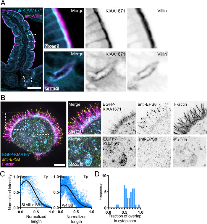
FIGURE 2:
KIAA1671 is an uncharacterized protein that targets the brush border cytoskeleton. (A) A paraffin-embedded mouse small-intestinal tissue section stained with anti-KIAA1671 (cyan) and anti-Villin (magenta) antibodies. Zoom 1 highlights apical staining on the villus, while Zoom 2 highlights apical staining in intestinal crypts. Scale bar = 50 µm. (B) Representative W4 cell expressing an EGFP-KIAA1671 construct (cyan), stained with an anti-EPS8 antibody (yellow) and phalloidin to mark F-actin (magenta). Zoom 1 highlights KIAA1671 at the base of the brush border, while Zoom 2 highlights KIAA1671 localization in the cytoplasm. (C) Linescans measuring anti-KIAA1671 (Left) or EGFP-KIAA1671 (Right) at the apical surfaces of intestinal tissue and W4 cells, respectively. n = 63 linescans of villus intestinal tissue from one independent staining; n = 40 line cans of W4 brush borders from three independent experiments. (D) Quantification of the fraction of overlap between KIAA1671 and F-actin. Bars indicate the fraction of total KIAA1671 signal that overlaps with F-actin signal. n = 40 cells from three independent experiments. All images are displayed as maximum-intensity projections.

