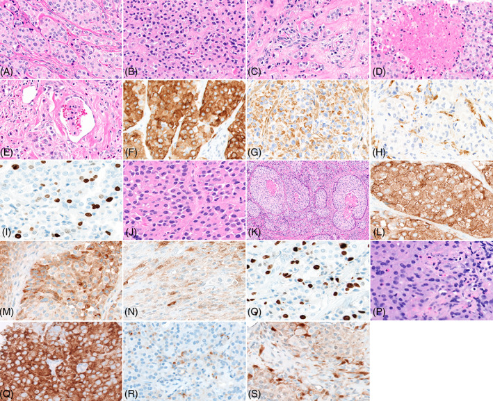
FIGURE 1.

Microscopic and immunohistopathological features of primary, recurrent and metastatic lesion. Panel A shows tumor cells organized in a conventional organoid pattern (40×). Panel B shows area of the lesion with a solid growth pattern (40×). Panel C shows cells with infiltrative growth pattern (40×). Panel D shows area of necrosis within the tumor (40×). Panel E shows area with lymphovascular invasion (40×). Panel F shows tumor cells with strong diffuse positive immunoreactivity to synaptophysin (40×). Panel G shows tumor cells with positive immunoreactivity to chromogranin (40×). Pane H shows sustentacular cells showing immunoreactivity to S100 (40×). Panel I shows reactivity of tumor cells to Ki67 (40×). Panel J shows tumor cells organized in a conventional organoid pattern (40×). Panel K shows area of necrosis (20×). Panel L shows tumor cells showing strong diffuse immunoreactivity to synaptophysin (40×). Panel M shows tumor cells showing diffuse immunoreactivity to chromogranin (40×). Panel N shows immunoreactivity of sustentacular cells to S100 (40×). Panel O shows reactivity of tumor cells to Ki67 (40×). Panel P shows histological features of tumor cells in metastatic lesion in lung (40×). Panel Q shows strong diffuse immunoreactivity to synaptophysin (40×). Panel R shows focal immunoreactivity of tumor cells to chromogranin (40×). Panel S shows immunoreactivity of sustentacular cells to S100 (40×).
