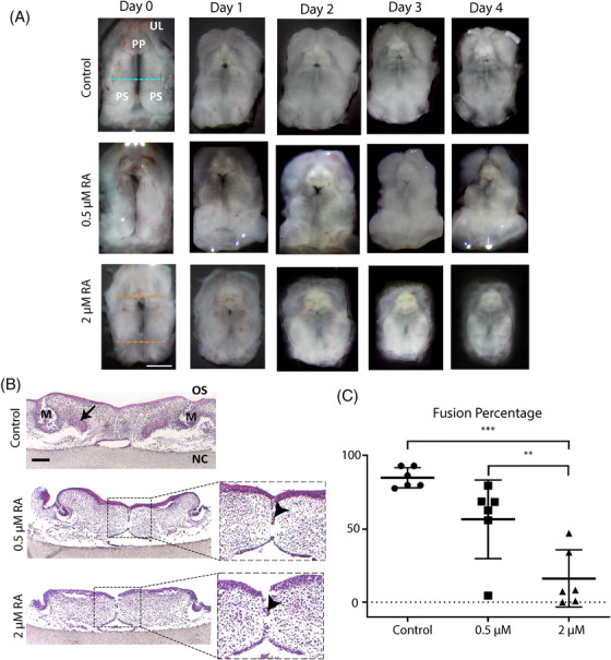
FIGURE 2.

Retinoic acid disrupts palate fusion in mouse palate cultures. Palates were isolated from E13.5 mouse embryos from nine different mothers, cultured oral‐side up for 4 days with 0.5–2 μM or without retinoic acid, fixed, and stained with HE. (A) Representative daily pictures of palates in culture. Green dotted line: middle region. Red dotted line: palate shelves length. PP: primary palate. UL: upper lip. PS: palate shelves. Scale bars: 1 mm. (B) HE staining of frontal sections. Representative pictures of the palates from the middle region, stained with HE. M: molar. OS: oral side. NC: nasal cavity. Arrowheads: medial epithelial seam. Scale bars: 200 μm. (C) Palate shelves fusion percentage. Six palates from each concentration group were used for the measurements. Ten consecutive sections from the middle of each palate were analyzed (* p < 0.05, *** p < 0.001)
