
Figure 4.
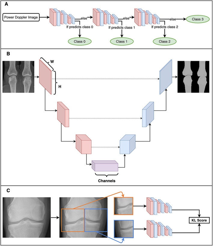
Three unique deep learning methods used in rheumatology. A, A cascade of convolutional neural networks (CNNs) used to classify power Doppler images. At each step, the CNN classifies the image as either a certain EULAR Outcome Measures in Rheumatology synovitis scoring class or any higher class (e.g., the first step classifies to either a class of 0 or >0). If the CNN determines that it belongs to a higher class, it is passed along to the next CNN, which performs the same task for the next highest class. Eventually, the final CNN simply classifies images as either class 2 or class 3. B, A simplified diagram of the U‐Net architecture (49). An image begins as an “N × N × C” shape, where “N × N” is the image size (e.g., 224 × 224 pixels) and “C” is the number of channels (typically 3 channels of red/green/blue for a color image). The model gradually reduces the size, while increasing the number of channels, until the bottom of the architecture is reached, and then the reverse occurs. Connections across the architecture (dashed lines) act as a “memory.” The image recovered at the end is a segmented image, partitioning the original into the relevant parts. In this example, the bones of 2 metacarpal joints are segmented from the plain radiograph. C, A single coronal radiograph of the knee joint split into 2 images: the right half of the knee and the horizontally flipped left half. Both images are passed through the same CNN before joining up to produce a Kellgren/Lawrence (K/L) composite score as the model output. Color figure can be viewed in the online issue, which is available at http://onlinelibrary.wiley.com/doi/10.1002/art.42296/abstract.