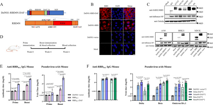
Fig. 1. Construction and evaluation of DelNS1-RBD4N-DAF live attenuated virus vaccine candidates.
A Illustration of construction of DelNS1-RBD4N-DAF. B Immunofluorescence (IF) assay of RBD expression with or without DAF in MDCK cells. MDCK cells were mock infected or infected with DelNS1, DelNS1-RBD or DelNS1-RBD-DAF viruses in the backbone of A/California/4/2009 (H1N1)16 at an MOI of 1 for 10 h and cells processed for IFA using antibodies specific for RBD (red). Nuclei were stained with DAPI (blue). Images are representative of three independent experiments. C Confirmation of N-glycosylation in RBD of DelNS1-RBD4N-DAF LAIV. MDCK cells were mock infected or infected with DelNS1-RBD (WT, lineage A virus), DelNS1-RBD with individual N-glycosylation mutations (A372T, G413N, D428N, or P521N), or DelNS1-RBD4N (4N) LAIV virus at an MOI of 1 for 10 h. Cell lysates were analyzed by western blot using anti-RBD antibody and anti-NP antibody. DelNS1-RBD, DelNS1-RBD4N, and DelNS1 LAIVs were also used to infect A549, BHK21 and MDCK cells to detect the expression of RBD. Images are representative of three independent experiments. D Schedule of immunization and blood collection for BALB/c mice. E Estimation of anti-RBD antibodies in mice prime-boost immunized intranasally with 2 × 10⁶ pfu of DelNS1-RBD-DAF with wild-type (WT) SARS-CoV-2 strain (lineage A) RBD (RBDWT-DAF, n = 4) or DelNS1-RBD with WT-RBD (RBDWT, n = 5) or DelNS1 vector (n = 5). At weeks 4 and week 6, blood was collected from mice and tested for anti-S1 RBD-specific IgG titers by ELISA assay and neutralization activity against pseudovirus expressing wild-type SARS-CoV-2 spike protein. F Estimation of anti-RBD antibodies in mice prime-boost immunized intranasally with 2 × 10⁶ pfu of DelNS1-RBD-DAF with Delta (B.1.617.2) RBD (Delta-DAF, n = 8) or Beta (B.1.351) RBD (Beta-DAF, n = 8), or DelNS1-RBD4N-DAF with Delta RBD4N (Delta4N-DAF, n = 8) or Beta RBD4N (Beta4N-DAF, n = 8), or DelNS1 vector (n = 8). At week 6, blood was collected from mice and tested for anti-S1 RBD-specific IgG titers and neutralization activity against pseudoviruses expressing spike proteins of SARS-CoV-2 variants. LOD: lower limit of detection. Error bars represent mean ± SD. Statistical analysis was performed using one-way ANOVA followed by Dunn’s multiple comparisons test: ****p < 0.0001, **p < 0.01, *p < 0.05, ns not significant. Numerical labels indicate fold difference. Mouse cartoon created with BioRender.com.

