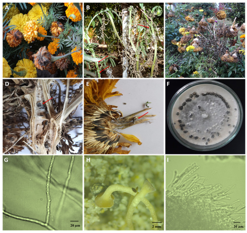
Figure 1.
Infected marigold plants and the isolated fungus. (A) Infected marigold plants showing white mycelium growth (indicated by arrows). (B) Infected flower buds showing white rot symptoms. (C) Severely infected garden showing infected, wilted, and dried plants. (D) Infected stem showing sclerotial development in the internal pith tissues (indicated by arrows). (E) Infected marigold flower with external and internal brown discoloration with embedded black sclerotia (indicated by arrows). (F) Pure culture of the infected fungus showing fluffy white mycelium and sclerotial ring. (G) Micrograph of the mycelium of Sclerotinia sclerotiorum isolated from marigold plants. (H) Induced formation of mature apothecia. (I) Rows of asci containing ascospores.

