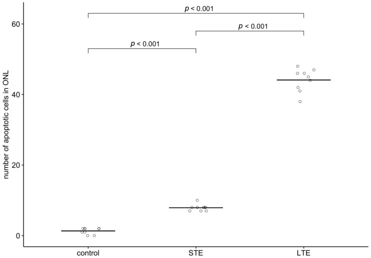
Figure 9.
Effect of blue light on the number of apoptotic cells in the outer nuclear layer in Wistar rat retinas. The rats were exposed to 12 h white light (150 lx) and 12 h darkness for 10 d (control, CTRL; 9 rats), cycles of 12 h blue light (150 lx) and 12 h darkness for 10 d (long-term exposure, LTE; 9 rats), and constant blue light (150 lx) for 2 d (short-term exposure, STE; 9 rats). Circles show the intensity of GFAP immunoreactivity in one retinal cryosection; horizontal lines show group means. p-values were calculated by quasi-Poisson regression followed by pairwise comparisons (Tukey’s method for p-value adjustment).

