Figure 10.
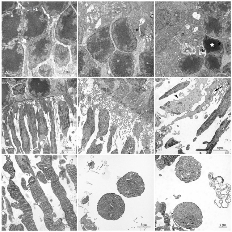
Effect of blue light exposure on the morphology of photoreceptors. Electron micrographs of retinas from Wistar rats. The rats were exposed to 12 h white light (150 lx) and 12 h darkness for 10 d (control, CTRL; 9 rats), cycles of 12 h blue light (150 lx) and 12 h darkness for 10 d (long-term exposure, LTE; 9 rats), and constant blue light (150 lx) for 2 d (short-term exposure, STE; 9 rats). Normal looking outer nuclear layers (ONL) in the (A) control and (D) STE groups. (G) ONL from the LTE group. Note: a single nucleus with pyknotic chromatin (star) present among normal looking nuclei in the ONL. Normal looking photoreceptor outer and inner segments from the (B) control and (E) STE groups. (H) Inner segments from the LTE group display some abnormalities. Note: some inner segments are thinner and longer (arrow), and some are swollen and thicker and have a lighter cytoplasm (arrowhead) than those in the control group. (C) Normal looking photoreceptor outer segments in the control group. In the (F) STE and (I) LTE groups, the apical parts of the photoreceptor outer segments form round or ellipsoidal structures containing tubules and vesicles.

