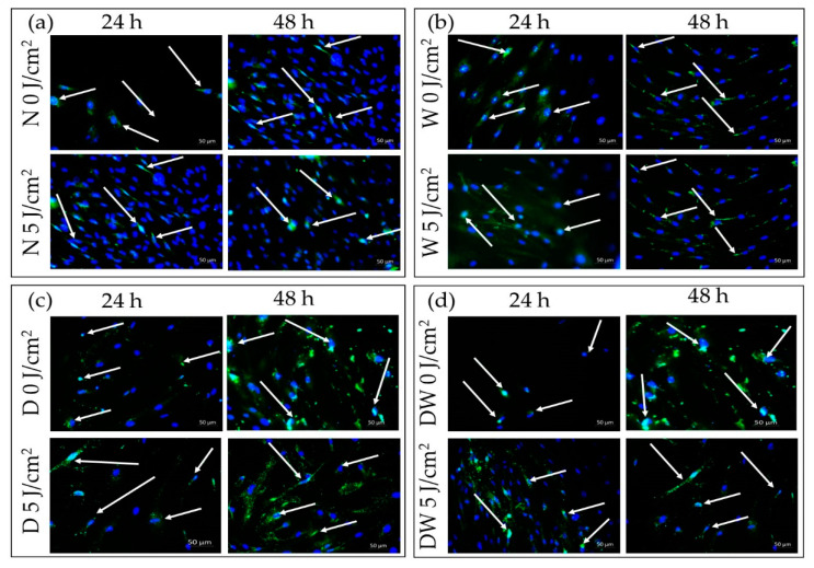
Figure 10.
Immunofluorescence images of phosphorylated (p-) ERK1/2 (MAPK) in non-irradiated and irradiated normal (N) (a), wounded (W) (b), diabetic (D) (c), and diabetic wounded (DW) (d) cells at 24 and 48 h after irradiation at 660 nm with 5 J/cm2. Phosphorylated protein appears green (FITC; green signals), while the nuclei appear blue (DAPI; blue signal); 200× magnification.

