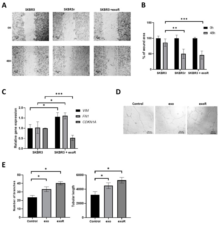
Figure 6.
Effects of exosomes in cell migration and angiogenesis. (A) Representative images of wound-healing cell migration assay of SKBR3, SKBR3r, and SKBR3 co-cultured with exosomes from SKBR3r (exoR). Magnification: 10×. (B) Quantification of the wound remaining area at 0 and 48 h. (C) Expression of EMT markers (VIM and FN1) and miR-146a-5p target CDKN1A, measured via RT-qPCR after co-culture of SKBR3 with exoR. GAPDH was used as endogenous mRNA gene, and the expression was calculated using the ΔΔCt method. (D) Representative images of in vitro angiogenesis assay of HUVEC-2 cells 8 h after addition of exo or exoR. Magnification: 5× (scale bar: 200 µm). (E) Representations of the number of branching points and the relative tubular lengths of HUVEC-2 cells 8 h after exo or exoR addition. Each experiment was performed in technical and biological triplicate. * p-value < 0.05; ** p-value < 0.01; *** p-value < 0.001. VIM: Vimentin; FN1: Fibronectin.

