Figure 7.
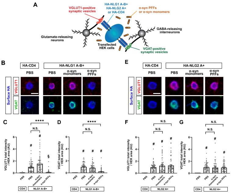
Co-culture-based artificial synapse formation assays reveal that α-syn PFFs diminish presynaptic differentiation induced by the NLG1 A-B+ isoform. (A) Schematic illustration of artificial synapse formation assays based on co-cultures of primary hippocampal neurons and HEK cells transfected with HA-NLG1/2 or HA-CD4 (a negative control). Co-cultured samples are immunostained for VGLUT1, an excitatory presynaptic vesicle protein, and VGAT, an inhibitory presynaptic vesicle protein, to visualize NLG-induced excitatory and inhibitory presynaptic differentiation, respectively. (B,E) Representative images from an artificial synapse formation assay showing the accumulation of VGLUT1 (red) and VGAT (green) induced by HA-NLG1 that lacks the splicing site A insert but possesses one at splicing site B (HA-NLG1A-B+; A) or by HA-NLG2 that possesses the splicing site A insert (HA-NLG2A+; D). HA-CD4 was used as a negative control. α-syn PFF treatment (400 nM, 24 h) dampens the presynaptic accumulation of VGLUT1 and VGAT induced by HA-NLG1A-B+ (B). In contrast, α-syn PFF treatment does not appear to affect HA-NLG2-induced accumulation of VGLUT1 or VGAT (E). Treatment with α-syn monomers has no significant effects on VGLUT1 or VGAT accumulation induced by HA-NLG1A-B+ or HA-NLG2A+. Scale bars: 20 μm. (C,D,F,G) Quantification of the presynaptic accumulation of VGLUT1 (C,F) and VGAT (D,G) in hippocampal neurons co-cultured with HEK293T cells expressing HA-NLG1A-B+ (C,D), HA-NLG2A+ (F,G), or HA-CD4, a negative control. Kruskal-Wallis one-way ANOVA, p < 0.0001. # p < 0.0001 and § p < 0.01 compared with HA-CD4 and **** p < 0.0001 for the indicated comparisons with PBS control by Dunn’s multiple comparisons test. N.S., not significant. Data are presented as mean ± SEM. (n > 60 cells for each from four independent experiments).

