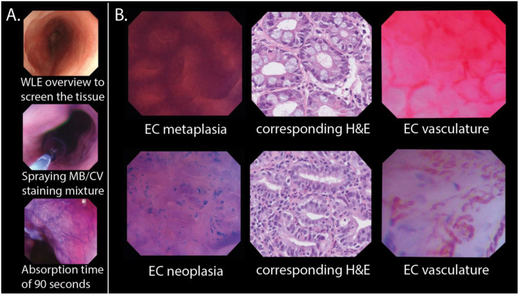
Figure 2.
In vivo EC procedure and classification. (A) Sequential assessment of the BE performed during endoscopic examination using EC. (B) Examples of EC metaplasia and EC neoplasia with their corresponding histopathology. Examples of EC images showing microvascular features in metaplastic and neoplastic tissue are shown to the right. The EC metaplasia and EC neoplasia images were used to create endocytoscopic BE datasets. BE: Barrett’s oesophagus; CV: crystal violet; EC: endocytoscopy; H&E: haematoxylin and eosin staining; MB: methylene blue; WLE: white light endoscopy.

