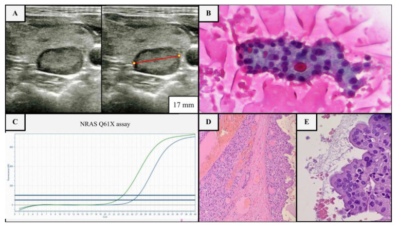
Figure 1.
Example of US, cytological and molecular assessment of a thyroid nodule in a 17-year-old female patient with multinodular goiter without clinical risk factors. (A) Nodular thyroid lesion of the right lobe, 17 mm in diameter (red line), isoechoic, with a mixed composition and smooth margins, no microcalcifications, deserving FNA according to ATA guidelines (≥1 cm partly solid nodule), but not deserving it according to ACR TI-RADS (class 2) (B) FNA shows a blood-rich background with fluid colloid, rare foamy histiocytes and numerous groups of thyroid cells mainly arranged in sheets and microfollicular structures, sometimes with oxyphilic appearance (class IV sec. TBSRTC, Papanicolau stained smear, ×40). (C) Molecular test (real time PCR) was performed, since the indeterminate cytology, starting from the needle wash, revealing the NRAS p.Q61X mutation (blue line mutated allele, green line wild type allele), a low-risk mutation group, which confirms the clonality of the lesion and its likely low biological aggressiveness. (D,E) Accordingly, a hemi-thyroidectomy was performed, revealing a partly cystic nodule composed of oncocytic thyrocytes, with pushing borders and a thin fibrotic capsule without invasion or lymphovascular infiltration, leading to the final histological diagnosis of NRAS-mutant oncocytic follicular adenoma (H & E, 10× and 40×).

