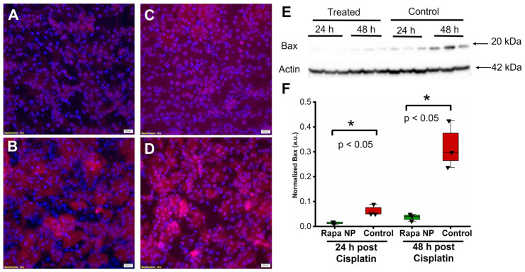
Figure 6.
Rapamycin PFC NP treatment reduced Bax expression in the kidney. (A,B) Representative images of Bax staining (red) in the kidney from mice 24 h after cisplatin injection with (A) or without (B) rapamycin PFC NP treatment. (C,D) Representative images of Bax staining (red) in the kidney from mice 48 h after cisplatin injection with (C) or without (D) rapamycin PFC NP treatment. DAPI nuclear staining is shown in blue. The size of the scale bar is 20 µm and the magnification is 40×. (E) Western blot of Bax showing rapamycin PFC NP treatment reduced Bax expression 48 h after cisplatin injection. β-Actin served as the loading control. (F) Quantification of Bax expression. (n = 3 per group, mean ± SEM).

