. 2023 Mar 22;14(15):4143–4151. doi: 10.1039/d3sc00886j

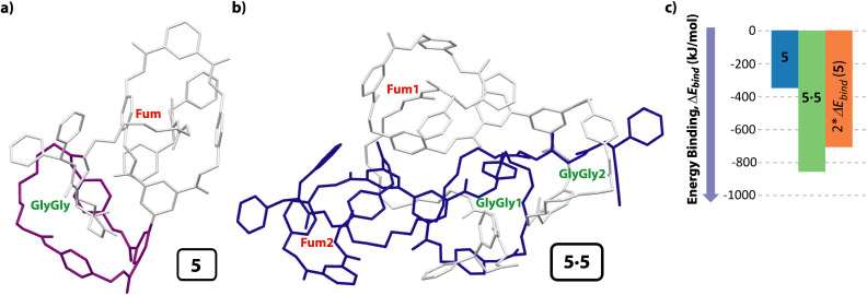
Fig. 5. Computational models at DFT level in CHCl3 (BP86-D3/def2-SVP) of: (a) [2]rotaxane 5; (b) cyclic hetero[4]pseudorotaxane (dimer 5·5); (c) diagram for visually comparing the binding energies of the monomer 5, the cooperatively self-assembled dimer 5·5 and that of the double of binding energy of the monomer 5. Both calculated structures are the lowest energy ones; ΔEbind: variation of the binding energy calculated as the difference between the energy of the complex minus the energy of all separated molecules that formed it (of the conformer with the lowest energy). For clarity the hydrogen atoms have been removed. Note that in the cyclic dimer 5·5, one of the molecules is colored in grey and the second one in dark blue. For clarity the diamide macrocycle is colored in purple in monomer 5.

Fig. 5