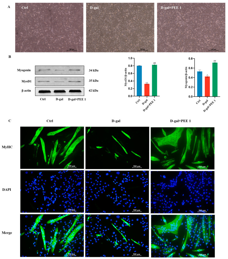
Figure 2.
PEE alleviated the impairment of differentiation capacity of D-gal-treated C2C12 cells. (A) Morphological changes in the number and size of myotubes after PEE treatment of C2C12 cells (scale bar = 100 μm). (B) Western blot and quantification for MyoD, and Myogenin. (C) The fluorescence staining for myosin heavy chain (MHC), indicator of Myotubular fusion (scale bar = 500 μm). All data are expressed as mean ± SEM (n = 3). * p < 0.05 and ** p < 0.01 vs. Ctrl; ## p < 0.01 vs. D-gal.

