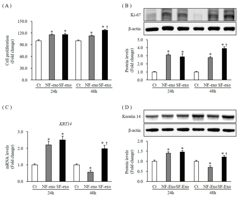
Figure 2.
Cell proliferation and expression of proliferation markers in normal human keratinocytes (NHKs) after NF- and HTSF-exosome treatment. NHK proliferation increased significantly at 24 and 48 h after treatment with 100 μg/mL NF- and HTSF-exosomes, respectively, compared with Dulbecco phosphate-buffered saline (DPBS)-treated control cells; there was a significant difference between NF- and HTSF-exosome treatment at 48 h (A). For the fold change, control cells were marked by a value of 100%. The cell proliferation assay was performed in triplicate. Protein expression of Ki-67 (B) and the mRNA and protein expression of keratin 14 (KRT14; C,D) in NHKs increased significantly at 24 and 48 h after treatment with 100 μg/mL HTSF-exosomes, except for NF-exosomes at 48 h, compared with that in the control cells. * p < 0.05 for NF- or HTSF-exosome-treated cells versus the corresponding matched control cells. † p < 0.05 for HTSF-exosome-treated cells versus NF-exosome-treated cells. Data represent the mean ± standard deviation (SD); n = 4. Ct, DPBS-treated control cells; NHK, normal human keratinocytes; NF-exo, normal tissue-derived fibroblast exosomes; SF-exo, hypertrophic scar tissue-derived fibroblast exosomes.

