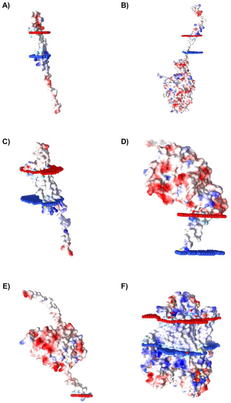
Figure 7.
Membrane regions’ spatial distribution and electrostatic potential determination of proteins identified for Caov-3 cell line. The proteins selected for Caov-3 cells were: (A) FXYD3, (B) ITGB2, (C) CLDN7, (D) ALPP, (E) SMPDL3B, and (F) STEAP4. For the spatial orientation, the red portion indicates the extracellular region, and the blue portion indicates the intracellular region. For the electrostatic potential determination, blue color indicates a positive charge and red color indicates a negative charge.

