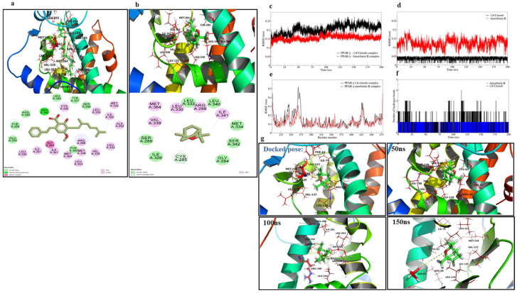
Figure 1.
Molecular docking and dynamics of 1,8-cineole with PPARγ. The docked pose and the interactions at the binding site of PPARγ. (a) Interactions of amorfrutin B and (b) interactions of 1,8-cineole. The analysis of the MD simulation (c) RMSD in PPARγ backbone atoms, (d) RMSD in 1,8-cineole atoms and amorfrutin B atoms, (e) RMSF in PPARγ residues, (f) hydrogen bond analysis for 1,8-cineole and amorfrutin B, (g) The analysis of trajectories extracted at different time intervals (50–150 ns) of the MD simulations.

