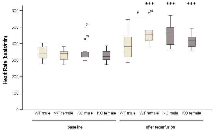
Figure 3.
In male wild-type (WT) mice, heart rate did not increase following ischemia/reperfusion. WT and cardiomyocyte-specific MAO-B knockout (KO) hearts were exposed to 45 min ischemia and 120 min reperfusion. Heart rate was measured baseline and after reperfusion. (WT male n = 14; WT female n = 9; KO male n = 11; KO female n = 11). Data are represented as box plots expressing median, 25% and 75 % quartiles, upper and lower whisker and outliers (○, ). * p < 0.05, *** p < 0.001 baseline vs. after reperfusion, Student’s t-test. Two-way ANOVA: p < 0.001, baseline vs. after reperfusion p < 0.001, genotype * sex p = 0.02.

