Figure 5.
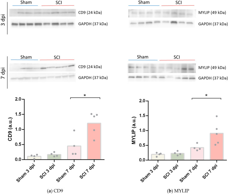
CD9 and MYLIP protein expression increase after spinal cord injury. Representative Western Blot images of CD9 (a) and MYLIP (b) with GAPDH as protein loading control at 3- and 7-days post-injury (dpi), each lane corresponds to an independent biological replicate. CD9 (a) and MYLIP (b) protein levels were quantified in sham and injured animals with a significant increase at 7 dpi for CD9 and MYLIP (n = 4–5). Data are expressed in relative intensity towards GAPDH, and bars represent the mean. * p < 0.05 versus sham, Student’s t-test.

