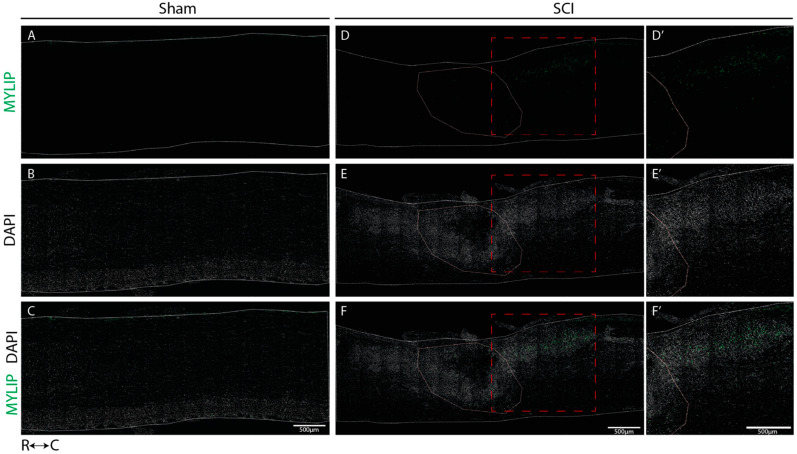
Figure 7.
MYLIP is expressed only after injury at the caudal side of the lesion. Representative images of sham (A–C) and injured spinal cords (D–F) demonstrate that MYLIP (green) is injury-induced with more prominent expression in the caudal region of the lesion (D’–F’) in all animals analyzed (n = 3). DAPI in grey. White dashed lines: spinal cord delimitation and core of the lesion; Red insert: close-up of the zone of interest R: rostral; C: caudal. Scale bar: 500 µm; 20× amplification.

