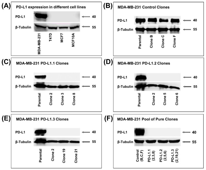
Figure 1.
PD-L1 knockout in MDA-MB-231 cell line using CRISPR/Cas9. (A) Western blot showing the level of PD-L1 in different breast cell lines. (B–E) Western blot showing the level of PD-L1 in the pure clones of the control, PD-L1.1, PD-L1.2, and PD-L1.3. (F) Western blot showing PD-L1 level in the pool of the three selected pure clones from the control and each design of sgRNA targeting PD-L1.

