Figure 3.
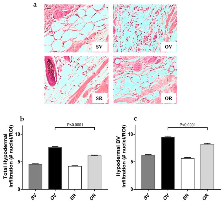
Decreased cellular infiltration in the hypodermis of mouse dorsal skins after a 7-day treatment with ovalbumin and resveratrol: (a) Hematoxylin and eosin (H&E) staining of skin tissues, focusing on the hypodermis after 7 days of epicutaneous exposure to saline (S) and vehicle for resveratrol (V), ovalbumin (O) and V, S with resveratrol (R), or O with R (scale bar = 50 mm). (b) Quantification of whole hypodermal cell infiltration after 7 days of epicutaneous exposure to S and V (dark grey filled bars), O and V (black filled bars), S with R (empty bars), or O with R (light grey bars). N = 6 mice per experimental group, 10 images per mouse, 10 ROI per image. (c) Quantification of hypodermal cell infiltration around blood vessels (BV) after 7 days of epicutaneous exposure to S and V (dark grey filled bars), O and V (black filled bars), S with R (empty bars), or O with R (light grey bars). #, number of, n = 6 mice, 10 images per mouse, 225–255 BV-containing ROI per experimental group.

