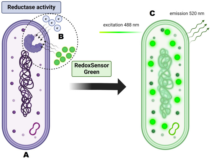
Figure 10.
Principles of RedoxSensorTM Green staining and its activity on the cellular level. (A) Analyzed cell is dyed with RedoxSensorTM Green. (B) Dye molecules permeate the cell membrane and interact with reductases. Reductase activity reflects changes in electron transport chain function and in vitality. (C) After excitation at 488 nm the dye emits green-fluorescent signal at 520 nm.

