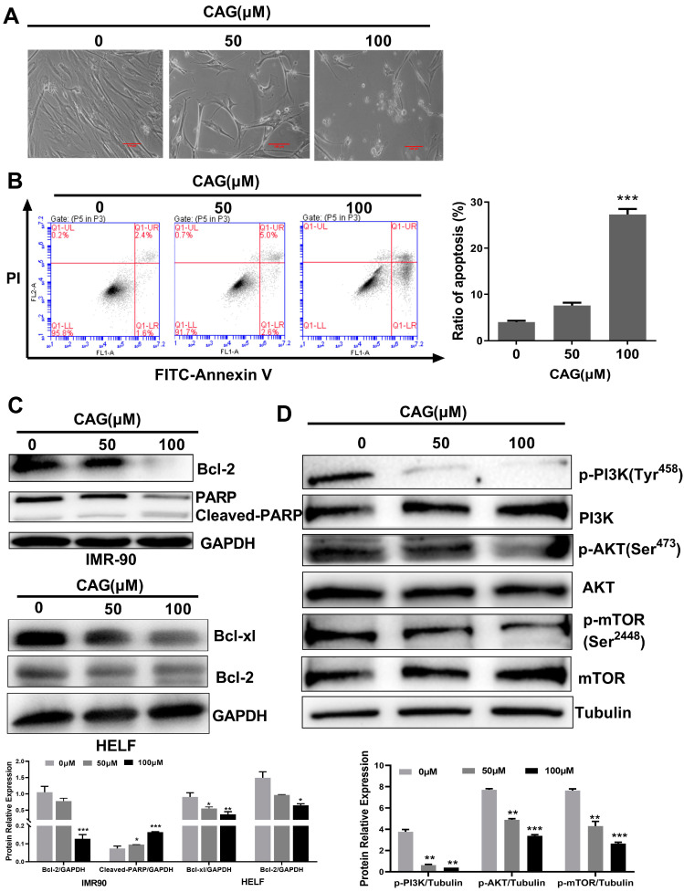
Figure 2.
CAG induces SCs apoptosis by inhibiting the antiapoptotic Bcl-2 family and PI3K/AKT/mTOR pathway. (A) Cell morphological changes in SCs after CAG treatment. Microscopic (magnification of 200×) images were taken. (B) Flow cytometric analysis of the cell death of SCs treated with CAG via annexin V/PI staining. (C,D) The expression levels of Bcl-2, PARP, Bcl-xl, and PI3K/AKT/mTOR pathway in SCs after incubation with indicated concentrations of CAG. Protein levels were determined using Western blots. The value represents the protein expressions compared to the GAPDH or Tubulin. * p < 0.05, ** p < 0.01, and *** p < 0.001.

