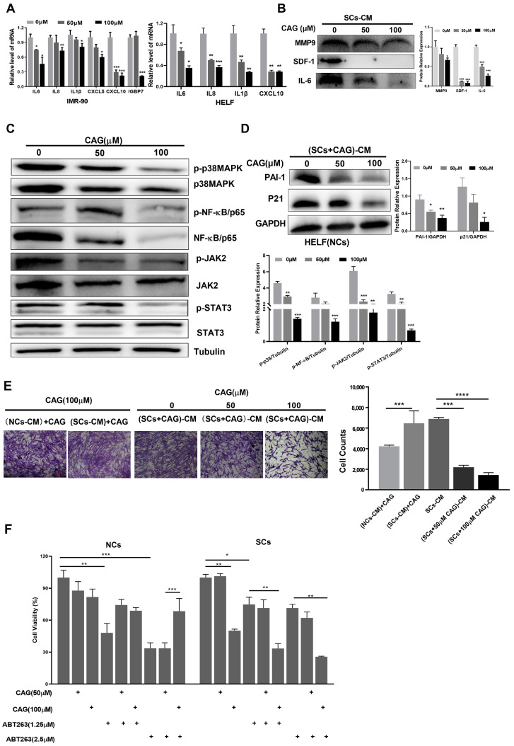
Figure 3.
CAG suppresses the SASP and decreases senescence and cell migration induced by SCs. (A) The relative mRNA abundance of key SASP components in senescent IMR-90 and HELF cells treated with different concentrations of CAG. RNA was collected and real-time PCR was performed. (B) CM was collected from senescent HELF cells (SCs-CM). Cytokine protein levels in CM were measured using Western blots. (C) The expression levels of p38, MAPK, NF-κB, JAK, and STAT3 in SCs after incubation with indicated concentrations of CAG. Protein levels were determined using Western blots. (D) NCs were treated for 48 h with CM collected from SCs which was pretreated with CAG. Expression levels of PAI-1 and P21 were determined using Western blots. (E) HELF cells were placed in inserts in the top chambers, and CM from different groups (SC-CM: CM collected from senescent HELF cells; (SC-CM) + CAG: CM collected from senescent HELF cells with added CAG; (SC + CAG)-CM: CM collected from senescent HELF cells which was pretreated with CAG) was introduced into the lower chambers. Cell migration was tested using crystal violet staining following 24 h of exposure. Microscopic (magnification of 200×) images were taken. Data are shown as the fold-change in cell number in each treatment vs. the SC-CM group. (F) HELF-VP16-induced senescent cells were treated with indicated concentrations of CAG and ABT263 for 48 h. Cell viability was assayed via MTT assay. * p < 0.05, ** p < 0.01, *** p < 0.001 and **** p < 0.0001.

