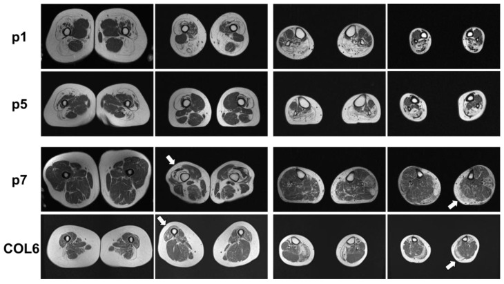
Figure 2.
Muscle MRI of p1, p5, and p7 as well as a patient with a pathogenic COL6A1 mutation. p1: Muscle MRI of p1 with known BICD2-associated myopathy showed fatty degeneration of the thigh muscles, pronounced in the lateral biceps femoris muscles (left panels) and the calf muscles, pronounced in the soleus and gastrocnemius muscles (right panels). p5: Muscle MRI of p5 with novel BICD2 and FLNC variants showed fatty degeneration of the vastus lateralis and medialis muscles as well as the lateral biceps femoris muscle in the thigh (left panels) and fatty degeneration of the gastrocnemius and soleus muscles (right panels). p7: Muscle MRI of p7 with novel BICD2 and COL6A1 variants showed distal fatty degeneration of the thigh muscles (especially vastus lateralis (left panels, white arrow)) and asymmetric, left, and distally pronounced fatty degeneration in the gastrocnemius muscle, caput mediale, soleus, and peroneus longus muscles (right panels, white arrow). COL6: Muscle MRI of a patient with COL6A1-associated myopathy shows typical distal atrophy of vastus lateralis muscle (left panels, white arrow) and gastrocnemius muscles (right panels, white arrow). White arrows indicate muscle degeneration typically associated with COL6A1-associated myopathy.

