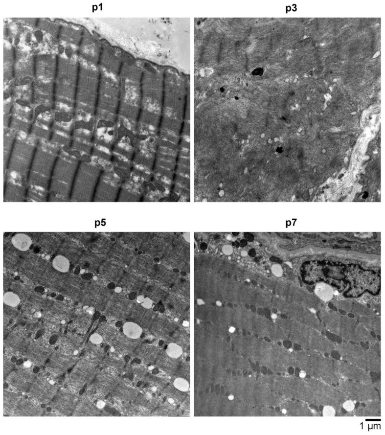
Figure 4.
Electron microscopy (EM) analysis of muscle biopsy material obtained from various affected individuals (p1 (p.Ser107Leu), p3 (p.Arg730His), p5 (p.Arg635Leu in BICD2 and p.Val758Met in FLNC), and p7 (p.Lys818Glu in BICD2 and p.Arg565Gln in COL6A1)) showed all had an advanced stage of myofibrillar breakdowns, lesions, and a massive presence of lysosomal vesicles, accompanied by enlarged polymorphic mitochondria and abundant autophagic vacuoles. In most myocytes, the sarcolemma appears with perforations and leaks, and the overall contractile apparatus shows only minor structural integrity. Scale bar: 1 μm.

